This lab manual provides essential guidance for students studying human anatomy and physiology, offering a structured approach to hands-on experiments and practical exercises. Designed for health-related programs, it enhances understanding through clear instructions, visual aids, and interactive activities, ensuring a comprehensive learning experience.
1.1 Purpose and Scope of the Lab Manual
The purpose of this lab manual is to provide a comprehensive, hands-on learning experience for students studying human anatomy and physiology. It is specifically designed for health-related programs, offering structured exercises and activities to enhance understanding of key concepts. The manual covers essential topics, including cellular structure, tissue types, skeletal and muscular systems, and physiological measurements. Its scope includes practical experiments, visual aids, and interactive tools to reinforce theoretical knowledge, ensuring students gain both conceptual and practical skills in anatomy and physiology.
1.2 Structure and Organization of the Manual
The lab manual is organized into clear, logical sections, each focusing on specific anatomical and physiological topics. It begins with an introduction, followed by essential equipment, cellular techniques, and systemic studies. Each chapter includes subheadings for detailed exercises, visual aids, and practical activities. The manual is designed to progress from basic to complex concepts, ensuring a gradual and comprehensive understanding. Its structure aligns with course objectives, making it easy for students to follow and apply their knowledge effectively.
1.3 Safety Protocols and Laboratory Etiquette
The manual emphasizes strict adherence to safety protocols to ensure a secure learning environment. Students are required to wear personal protective equipment, such as gloves and goggles, when handling biological specimens or chemicals. Proper handling and disposal of biohazardous materials are clearly outlined. Emergency procedures, including spill management and injury response, are detailed to prepare students for unforeseen situations. Additionally, the importance of maintaining a clean workspace and respecting shared equipment is stressed to promote a culture of responsibility and cooperation in the lab setting.
Essential Laboratory Equipment and Tools
This section introduces the fundamental equipment used in anatomy and physiology labs, including microscopes, dissection tools, and physiological measurement devices, ensuring students are familiar with their proper use and maintenance.
2.1 Microscopes and Their Accessories
Microscopes are essential tools for studying cellular structures in anatomy and physiology labs. Common types include compound and stereo microscopes, each with specific accessories like eyepieces, objective lenses, and stages. Proper use involves focusing techniques and maintaining cleanliness to ensure clarity. Accessories like immersion oil and slides enhance imaging quality. Understanding microscope operation is crucial for accurate observations, making them indispensable for detailed tissue and cell examinations in laboratory settings.
2.2 Dissection Tools and Supplies
Dissection tools and supplies are crucial for exploring anatomical structures in physiology labs. Essential tools include scalpels, forceps, dissecting scissors, and probes. These instruments allow precise dissection and examination of tissues and organs. Additional supplies like dissecting trays and storage containers help maintain organization and safety. Proper use and maintenance of these tools are vital for effective learning and experimentation, ensuring a comprehensive understanding of human anatomy and physiology through hands-on practice.
2.3 Physiological Measurement Devices
Physiological measurement devices are essential tools for assessing bodily functions in anatomy and physiology labs. Common devices include electrocardiograms (ECG) for heart activity, spirometers for lung capacity, and blood pressure monitors. These tools provide quantitative data, enabling students to observe and analyze physiological responses. Proper use of these devices enhances understanding of human body functions, allowing for accurate measurements and interpretations of physiological processes during experiments and studies.
Cellular and Histological Techniques
This section explores methods for preparing and examining cells and tissues, including slide preparation, staining, and microscopy. These techniques are crucial for understanding cellular structure and function.
3.1 Preparing and Staining Microscope Slides
Preparing and staining microscope slides involves several key steps to ensure clear visualization of cellular structures. Begin by cleaning and labeling slides, then cut thin tissue sections using a microtome. Place sections on slides and allow them to air-dry. Staining is performed using dyes like hematoxylin and eosin to differentiate cell components. Fixation and dehydration steps are critical for preserving tissue integrity. Proper technique ensures high-quality samples for microscopy, enabling accurate observations of cellular details and structures. This process is fundamental for histological analysis in anatomy and physiology studies.
3.2 Identifying Cellular Structures Under a Microscope
Identifying cellular structures under a microscope involves observing and recognizing key components such as the nucleus, mitochondria, and cytoplasm. Proper use of the microscope, including focusing and adjusting magnification, is essential for clear visualization. Staining techniques enhance contrast, allowing differentiation of cellular features. Students learn to distinguish between various organelles and their functions, gaining insights into cellular organization. This skill is fundamental for understanding histological samples and diagnosing abnormalities, making it a critical component of anatomy and physiology lab training.
3.3 Understanding Tissue Types and Their Functions
Understanding tissue types involves studying their structure, function, and role in the body. The four primary tissue types—epithelial, connective, muscle, and nervous—each serve distinct functions. Epithelial tissues form barriers and line surfaces, while connective tissues provide support and connectivity. Muscle tissues enable movement, and nervous tissues facilitate communication through electrical signals. Lab exercises involve identifying these tissues under a microscope and exploring their contributions to overall physiology. This knowledge is essential for understanding how tissues interact to maintain homeostasis and support organ systems.
Skeletal and Muscular System Exercises
This section focuses on hands-on activities to identify bones, joints, and muscle groups, enhancing understanding of their structure and function. Practical exercises include mapping muscles and observing joint movements, providing a comprehensive exploration of the skeletal and muscular systems.
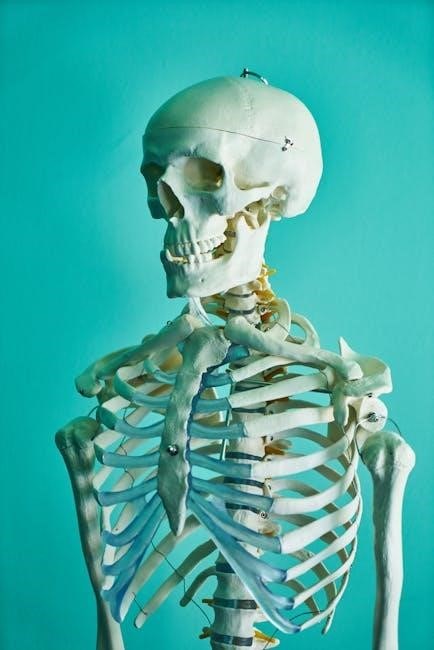
4.1 Identification of Bones and Joints
This exercise focuses on the detailed identification and classification of human bones and joints. Students learn to distinguish between various bone types, such as long, short, flat, and irregular bones, using detailed charts and 3D models. The activity emphasizes understanding joint structures, including synovial, cartilaginous, and fibrous joints, through hands-on examination of prosected specimens or anatomical models. This practical approach enhances recognition of anatomical landmarks and improves comprehension of skeletal system organization and functionality.
4.2 Mapping Muscle Groups and Their Functions
This section guides students in identifying and mapping major muscle groups, focusing on their anatomical locations and physiological functions. Through hands-on activities, such as palpation and model exploration, students learn to associate muscles with their actions, like flexion, extension, and rotation. Practical exercises emphasize understanding muscle group synergies and antagonisms, enhancing comprehension of human movement and its application in clinical and therapeutic contexts.
4.3 Observing Joint Movements and Biomechanics
This section focuses on analyzing joint movements and understanding biomechanical principles. Students explore types of joints, such as synovial and cartilaginous, and their range of motion. Through palpation and model examination, learners observe how bones articulate and function during activities like flexion, extension, and rotation. Practical exercises emphasize understanding movement patterns, forces, and muscle interactions, providing insights into human locomotion and its relevance in fields like physical therapy and rehabilitation.
Nervous System and Sensory Organs
This section explores the structure and function of the nervous system, including the brain, spinal cord, and peripheral nervous system. It also covers sensory organs like the eye and ear, focusing on how they detect stimuli and transmit signals. Hands-on activities include nerve reflex testing and anatomical examinations to understand neural pathways and sensory function, preparing students for advanced studies in neurology and rehabilitation.
5.1 Exploring the Brain and Spinal Cord
This section provides a detailed examination of the brain and spinal cord, focusing on their structural and functional components. Students will identify key brain regions, such as the cerebrum, cerebellum, and brainstem, and explore their roles in controlling voluntary and involuntary functions. The spinal cord’s role in nerve transmission and reflex actions is also highlighted. Activities include dissection, histological slide analysis, and anatomical model studies to visualize neural pathways and understand the central nervous system’s integration of sensory and motor functions.
5.2 Testing Reflexes and Sensory Responses
This section focuses on assessing reflex actions and sensory responses to understand neural pathways. Students perform tests such as patellar reflex and pain perception using tools like a reflex hammer and sensory response kits. Analysis involves documenting responses and correlating them with nervous system functions. These tests are crucial for diagnosing neurological conditions. This exercise enhances understanding of the nervous system’s role in involuntary and voluntary actions.
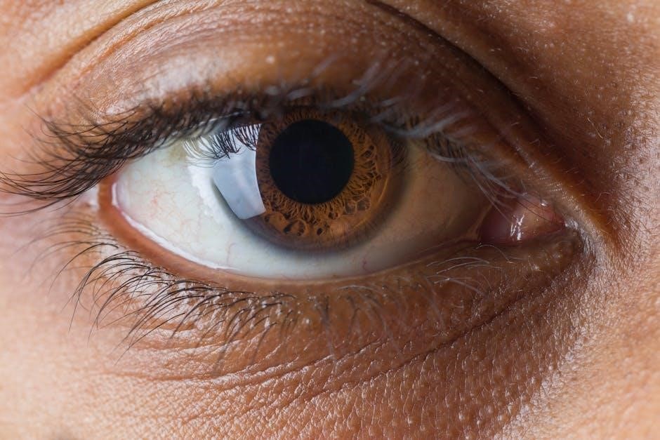
5.3 Investigating the Structure of the Eye and Ear
This section involves examining the anatomical structures of the eye and ear to understand their functions. Through dissection or models, students identify components like the cornea, lens, retina, and eardrum. Observations focus on how these parts contribute to vision and hearing. The exercise aids in understanding sensory mechanisms and the relationship between structure and function, essential for diagnosing disorders and clinical applications.

Cardiovascular and Respiratory System Labs
This section explores the heart, blood vessels, and lungs through hands-on activities. Students analyze blood components, measure heart rate, and study breathing mechanisms to understand circulation and gas exchange.
6.1 Analyzing Blood Smears and Blood Types
This exercise involves preparing and examining blood smears under a microscope to identify blood cells, including red blood cells, white blood cells, and platelets. Students learn to differentiate cell types based on size, shape, and staining characteristics. Blood typing is also explored, focusing on the ABO and Rh blood group systems. The process includes testing for antigens and antibodies to determine blood type, understanding cross-matching, and discussing the clinical significance of blood compatibility in transfusions. Clear instructions and safety protocols are emphasized for accurate results.
6.2 Measuring Heart Rate and Blood Pressure
This section introduces techniques to measure heart rate and blood pressure, essential for assessing cardiovascular health. Students learn to palpate radial or carotid pulses to determine heart rate in beats per minute (bpm). Blood pressure is measured using a sphygmomanometer, emphasizing proper cuff placement and recording systolic and diastolic pressures. The exercise explores how physical activity, stress, or posture affects these measurements, while ensuring accurate data recording and adhering to safety protocols for reliable results.
6.3 Understanding Lung Capacity and Gas Exchange
This section explores respiratory physiology through experiments measuring lung capacity and analyzing gas exchange. Students use spirometers to record vital capacity, tidal volume, and inspiratory/expiratory reserve volumes. Activities demonstrate how oxygen and carbon dioxide exchange occurs in alveoli, emphasizing the role of diffusion gradients. Factors influencing lung function, such as health conditions or physical activity, are discussed, with data interpretation linking anatomical structures to physiological processes, ensuring a comprehensive understanding of respiratory mechanics and their clinical relevance.

Digestive and Urinary System Experiments
This section involves hands-on experiments exploring digestive enzyme activity, pH levels, and urinary system functions, providing practical insights into their physiological roles and processes.
7.1 Examining the Structure of the Digestive Tract
This exercise involves the microscopic examination of histological slides to identify the layers of the digestive tract, including the mucosa, submucosa, muscularis, and serosa. Students learn to distinguish between different regions, such as the esophagus, stomach, small intestine, and large intestine, by observing structural adaptations like villi, microvilli, and glandular tissue. This hands-on activity enhances understanding of how the digestive tract’s anatomy correlates with its physiological functions, such as absorption, digestion, and waste elimination.
7.2 Testing Enzyme Activity and pH Levels
This lab involves testing the activity of digestive enzymes and measuring pH levels in different regions of the digestive tract. Students use pH indicators to determine acidity in gastric and intestinal juices. Enzyme activity is assessed using colorimetric tests, observing how pH affects enzyme effectiveness. The exercise demonstrates how specific enzymes, like pepsin and amylase, function optimally at particular pH levels. This hands-on approach clarifies the relationship between pH, enzyme activity, and digestive processes, preparing students for real-world applications in physiology and clinical settings.
7.3 Understanding Kidney Function and Urinalysis
This section explores the role of kidneys in filtering blood, regulating electrolytes, and excreting waste. Students perform urinalysis to examine urine components, including pH, protein, and glucose levels. The lab involves testing samples with dipsticks and microscopy to detect abnormalities. By analyzing these findings, students understand kidney function and its impact on overall health. This exercise prepares them to identify potential renal issues and interpret urinalysis results, reinforcing their knowledge of urinary physiology and clinical diagnostics.
Endocrine and Reproductive System Investigations
This section explores the structure and function of endocrine glands, hormones, and reproductive systems. Practical investigations include identifying glands, examining reproductive organs, and analyzing hormonal processes.
8.1 Identifying Endocrine Glands and Their Hormones
This section focuses on the identification and function of major endocrine glands, such as the pituitary, thyroid, adrenal, pancreas, and gonads. Students learn to locate these glands in anatomical diagrams and associate them with their respective hormones. Practical exercises involve distinguishing between endocrine and exocrine glands, understanding hormone production, and exploring the role of hormones in regulating bodily functions. Hands-on activities and visual aids enhance comprehension of endocrine physiology and its significance in maintaining homeostasis.
8.2 Exploring the Male and Female Reproductive Systems
This section provides a comprehensive study of the male and female reproductive systems, focusing on their anatomical structures and physiological functions. Hands-on activities include identifying reproductive organs, comparing male and female systems, and understanding the role of hormones in reproductive processes. Students also explore the relationship between the endocrine system and reproductive health, as well as common disorders and their implications. This exercise prepares future healthcare professionals to understand the intricacies of human reproduction and associated clinical conditions.
8.3 Observing Fetal Development Stages
This section guides students through the exploration of human fetal development, emphasizing key stages from conception to birth. Hands-on activities include examining models and digital simulations to visualize growth milestones. The lab focuses on identifying critical developmental phases, such as organogenesis and sensory system maturation. Students also analyze factors influencing fetal growth and potential abnormalities. This exercise enhances understanding of prenatal physiology and prepares learners to recognize normal vs. abnormal development patterns in clinical settings.
Clinical Applications and Case Studies
This section connects lab findings to real-world clinical scenarios, enabling students to apply anatomical and physiological knowledge to diagnose and manage medical conditions through case-based learning.
9.1 Applying Lab Findings to Real-World Scenarios
This section bridges the gap between laboratory experiments and practical medical applications. Students learn to interpret lab results in clinical contexts, such as diagnosing conditions or monitoring treatments. By analyzing case studies, learners develop critical thinking and problem-solving skills, essential for patient care. Real-world examples, like interpreting blood pressure readings or urinalysis results, are used to illustrate how lab data informs medical decisions. This application enhances understanding of physiological processes and prepares students for clinical practice.
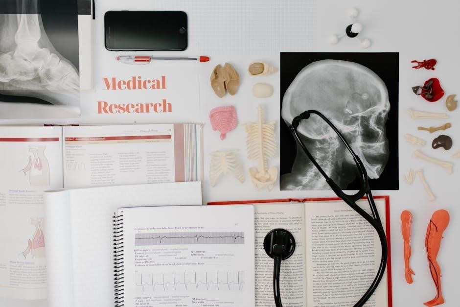
9.2 Analyzing Medical Imaging Techniques
Medical imaging techniques, such as X-rays, MRIs, and CT scans, are essential tools for visualizing internal structures. This section teaches students to interpret imaging results, identifying anatomical landmarks and pathological changes. Hands-on activities involve correlating images with lab findings to enhance diagnostic skills. Students explore how imaging aids in detecting fractures, tumors, or organ dysfunction, preparing them to apply these skills in clinical settings for accurate diagnoses and effective treatment planning;
9.3 Solving Case Studies Using Lab Knowledge
This section equips students with the skills to apply lab-derived knowledge to real-world clinical scenarios. Through detailed case studies, students analyze symptoms, interpret lab data, and correlate findings with anatomical and physiological principles. Activities focus on diagnosing conditions, understanding treatment options, and predicting outcomes. By integrating lab insights with patient histories, students refine their critical thinking and problem-solving abilities, preparing them for effective decision-making in healthcare settings.
Lab Safety and Biohazard Management
This section emphasizes the importance of maintaining a safe lab environment, proper handling of biohazardous materials, and adhering to emergency protocols to protect students and staff.
10.1 Handling Biohazardous Materials Safely
Proper handling of biohazardous materials is critical to prevent exposure and contamination. Use personal protective equipment (PPE) such as gloves and lab coats. Ensure materials are stored in sealed, labeled containers and disposed of in designated biohazard waste bins. Decontaminate surfaces and tools after use, and follow specific protocols for spills or accidents. Always wash hands thoroughly after handling biohazardous substances. Adhere to laboratory safety guidelines to maintain a secure environment for all personnel involved.
10.2 Proper Use of Personal Protective Equipment
Personal Protective Equipment (PPE) is essential for safeguarding against lab hazards. Always wear lab coats, gloves, goggles, and face masks when handling biohazardous materials or chemicals. Ensure PPE fits correctly and is free from damage. Use disposable gloves for direct contact with specimens and replace them immediately if torn. Remove PPE carefully to avoid contamination, especially when handling infectious agents. Properly dispose of used PPE in designated bins. Adhering to PPE protocols protects against chemical splashes, biological exposure, and sharp object injuries, ensuring a safer laboratory environment.
10.3 Emergency Procedures in the Lab
In case of emergencies, evacuate the area immediately and alert others. Use fire extinguishers for small fires and ensure exits are clear. For chemical spills, wear PPE, contain the spill, and neutralize if trained. In case of exposure, flush skin or eyes with water and seek help. For injuries, apply first aid and report incidents. Keep emergency contact numbers accessible. Regular drills ensure preparedness. Always follow lab-specific protocols and remain calm to manage situations effectively, prioritizing safety and minimizing risks. Proper training is essential for handling emergencies efficiently.
Lab Report Writing and Documentation
This section guides students in structuring lab reports, recording observations, and interpreting results. It emphasizes clarity, conciseness, and proper documentation to ensure accurate and professional presentation of findings.
11.1 Structuring a Lab Report
A well-structured lab report includes a clear title, introduction, materials list, procedures, results, discussion, and references. The title should concisely state the experiment’s purpose. The introduction provides context and objectives. Materials and procedures detail what was used and done. Results present data objectively, while the discussion interprets findings, linking them to broader concepts. Proper formatting ensures clarity and professionalism, making the report accessible and informative for readers.
11.2 Recording Observations and Data
Accurate and detailed recording of observations and data is crucial for reliable lab results. Use standardized tools like charts, graphs, and tables to organize findings. Note measurements, visual observations, and any unusual occurrences. Include timestamps and experimental conditions to ensure reproducibility. Avoid subjective interpretations; stick to factual data. Use cameras or sketches for visual documentation. Ensure consistency in units and formatting. Double-check data for accuracy before finalizing. This step forms the foundation for meaningful analysis and interpretation in later stages of the lab report.
11.3 Interpreting and Presenting Results
Interpreting and presenting results effectively communicates lab findings. Use clear graphs, tables, and captions to illustrate key data. Ensure conclusions align with objectives and hypotheses. Highlight trends and correlations while addressing anomalies. Avoid unsupported assumptions. Present data concisely, using visual aids to enhance understanding. Compare results with expected outcomes and discuss implications. Accurate and well-organized presentation ensures clarity and credibility, making it easier for readers to grasp the significance of the experiments and their relevance to human anatomy and physiology concepts.
Review of Key Anatomical and Physiological Concepts
This section summarizes major anatomical systems, such as skeletal, muscular, nervous, and cardiovascular, emphasizing their structural and functional integration. It highlights key physiological processes and common lab errors, providing practical troubleshooting tips to enhance understanding and lab performance.
12.1 Summary of Major Body Systems
This section provides a concise overview of the human body’s primary systems, including skeletal, muscular, nervous, cardiovascular, respiratory, digestive, urinary, endocrine, and reproductive. Each system’s structure and function are summarized, emphasizing their roles in maintaining homeostasis and overall health. Key physiological processes, such as blood circulation, nerve signaling, and digestion, are highlighted to reinforce understanding of how these systems interact to sustain life and enable bodily functions.
12.2 Integration of Structure and Function
This section explores how the structure of anatomical components directly relates to their physiological functions. It emphasizes the interconnectedness of body systems, illustrating how each system’s design enables its specific role in maintaining homeostasis. Through detailed lab exercises, students examine how structures like bones, muscles, and organs function together to support movement, circulation, and overall bodily processes, reinforcing the principle that form and function are inherently linked in human anatomy and physiology.
12.3 Common Lab Mistakes and Troubleshooting
This section highlights frequent errors students make during lab exercises, such as incorrect microscope focusing or improper slide preparation. It provides practical solutions, like recalibrating instruments or re-staining samples. Troubleshooting tips address issues like blurry observations or misidentified structures, ensuring accurate results. By understanding common pitfalls, students can refine their techniques and improve lab outcomes, fostering a more efficient and effective learning experience in anatomy and physiology labs.